 Increasing the mA and lowering the publicity time by a proportionate amount results in a radiograph much less likely to be degraded by movement. As a rule, it is best to attenuate the exposure time but keep an acceptable mAs and scale of contrast. Increasing kVp increases the number of photons penetrating the subject and so darkens the image. This effect can be used inside limits to correct an underexposure. This is an entire system with no further equipment wanted to be purchased in order to image animal patients.
 Sound waves sent into the physique are reflected off an internal tissue interface. Hundreds of those mirrored alerts create an image of the organ, which could be visualized on the ultrasound machine monitor. Nuclear medicine is an imaging approach that involves the injection of a radionuclide and then monitoring the distribution and intensity of the radioactivity throughout the body with a gamma camera. The course of is very delicate to abnormalities in the bone and can be used to evaluate the liver, kidneys, and thyroid. In addition, the Radiology Service offers I-131 therapy for therapy of hyperthyroidism in cats. Magnetic Resonance Imaging (MRI) is an imaging method that uses highly effective magnets to align hydrogen atoms and radiofrequency waves to systematically alter this alignment.
Refurbished DR Tech 8×10 Flat Panel System
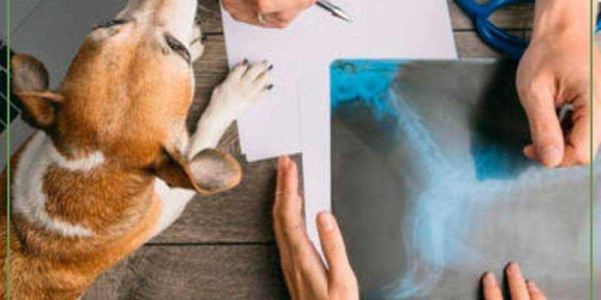
Radiographic examinations have to be carried out with correct respect for radiation security procedures. Diagnostic x-ray machines are potent sources of radiation and might, if improperly used, lead to injurious publicity to personnel over time. The publicity elements used in trendy x-ray systems are substantially decrease than these used prior Laboratorio de exames animais to now however can still end in damage. It is rarely acceptable to carry animals without the use of lead-impregnated aprons and gloves to lower publicity to the arms and physique of personnel from scattered radiation. Leaded gloves shouldn't be used throughout the main beam of the x-ray machine. These gloves and aprons scale back exposure from scatter radiation by an element of ~1,000 but reduce publicity from the first beam by only an element of ~10. Upper limb, cervical backbone, and skull research in horses are significantly prone to lead to substantial publicity of the upper physique and head to anyone holding the film/detector or the horse.
If used properly, this technique leads to practically similar image exposures between animals. However, acceptable kV settings are wanted, and constant animal positioning is critical. Identical positioning between animals is required to attain identical pictures. Placing the guts or lungs over the AEC sensor leads to radically different radiographs. AEC might be most effective when large numbers of pictures are being accomplished of the identical anatomic area by the identical personnel. AEC is usually not utilized in most veterinary applications because of the extensive variation in body sizes and conformation of canine. Increasing the mA setting on the machine increases the variety of x-rays produced.
Portable X-Ray
This can be prevented by taking a few extra seconds to properly place the animal for the first image. Individuals concerned in taking radiographic images ought to be monitored for radiation publicity. This is crucial to identify and proper circumstances that can lead to excessive radiation publicity to personnel. Monitoring of exposure also offers evidence of correct adherence to radiation safety requirements if questions come up as to whether an employee’s medical situation could be related to radiation publicity. Several corporations present this service for a relatively nominal fee. The difference between the 2 techniques lies in the intermediate step of exposing a plate in CR, which is then placed in a reader.
Our radiology group, a lot of whom are board-certified, has advanced coaching and experience that enables them to see abnormalities and make clear findings using photographs created by way of diagnostic imaging procedures. Using these images, we work with your liked ones veterinarian and other MedVet specialists to diagnose and Laboratorio de exames animais develop a therapy plan on your pet. Radiographic exposure of movie alone lacks sufficient contrast to evaluate many buildings; therefore, distinction procedures are used to extend the native distinction of organs and lesions, to separate them from surrounding tissues. Contrast procedures have been developed to extend the native distinction of organs and lesions and to judge the operate of some organs such because the GI tract. The portability of digital photographs and the speed and value of the internet has led to much greater access by veterinarians in non-public apply to the interpretive expertise of radiologists and different specialists. This has the potential to improve the standard of veterinary practice worldwide, not solely in the subject of imaging but in lots of different specialties. Processing algorithms are important to the event of diagnostic pictures.
People Results